Xiaojun Lian · Pennsylvania State University · PhdFit
Resume-aware faculty matching
Find professors who actually fit you
Upload your resume. PhdFit's six research agents compare your background with faculty profiles, recent publications, lab focus, and outreach opportunities, then rank professors with evidence you can review.
Resume-aware matchingEvidence from recent researchOutreach drafts included
Advisor Match Brief Resume uploaded
Resume signals5 found
NLPHCIFairnessGraph MLPyTorch
Six-agent analysisRunning
Axel · Candidate AnalystResume parsed
Nova · Professor ResearcherRecent papers
Echo · Match ExplainerEvidence linked
Atlas · Strategy AdvisorFit ranked
Lyra · Outreach WriterDraft queued
Juno · Meeting Prep CompilerNext step ready
SC
Dr. Sarah Chen
Stanford · NLP & interpretability
91
fit
Fairness + NLP projects overlap with her recent work.
Strong methods fit: model evaluation and interpretability.
Clear outreach angle for current trustworthy AI research.
Outreach angle
Ask how her lab is extending interpretability methods into fairness audits for real-world AI systems.
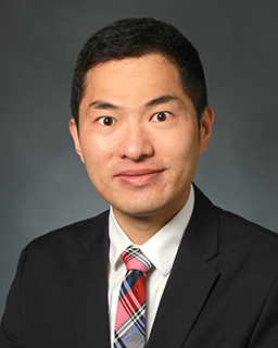
Xiaojun Lian
· Associate Professor of Biomedical EngineeringVerified
Pennsylvania State University · Biomedical Engineering
Xiaojun Lian is an Associate Professor of Biomedical Engineering at Penn State University. His research focuses on cell and molecular bioengineering, with particular emphasis on stem cell differentiation, cardiovascular regeneration, and bioelectronic interfaces. He has contributed to the development of methods for programming human pluripotent stem cells into various cell lineages, including neutrophils, endothelial progenitors, and cardiomyocytes, utilizing chemically defined media and genome editing techniques. His work also involves engineering bio-optoelectronic stimulators for cardiac applications and advancing genome and RNA editing in human pluripotent stem cells. Dr. Lian's research aims to address challenges in regenerative medicine and develop innovative bioengineering solutions for tissue repair and disease modeling.
The human heart, originating from the splanchnic mesoderm, is the first functional organ to develop, co-evolving with the foregut endoderm through reciprocal signaling. Previously, cardioid models offered new insights on cardiovascular cell lineages and tissue morphogenesis during heart development, while mesoderm-endoderm crosstalk remain incompletely understood. Here, we integrated micropatterned cardioids, CRISPR-engineered reporter hiPSCs, deep-tissue imaging, and single-cell RNA sequencing (scRNA-seq) to explore synergistic mesoderm-endoderm co-development. scRNA-seq with PHATE trajectory mapping reconstructed lineage bifurcations of mesoderm-heart and endoderm-foregut lineages, identifying key cell types in cardiac and hepatic development. Ligand-receptor interaction analysis highlighted mesodermal cells enriched in non-canonical WNT, NRG, and TGF-β signaling, while endodermal cells exhibited VEGF and Hedgehog activity. We found that micropattern sizes influenced cellular composition, cardioid cavitation, contractile functions, and mesoderm-endoderm signaling crosstalk. The cardioids generated from 600 µm diameter circle patterns showed larger cavity formation resembling early heart chamber formation. Our findings establish micropatterned cardioids as a model for mesoderm-endoderm co-development, enhancing our understanding of heart-foregut synergy during early embryogenesis.
Genetically encoded imaging reporters are critical tools for tracking cell fate and function in regenerative medicine. Gas vesicles (GVs), air-filled protein nanostructures derived from bacteria, offer unique advantages for noninvasive imaging due to their acoustic and optical properties. In this study, we engineered human pluripotent stem cells (hPSCs) to express GVs using a doxycycline (Dox)-inducible system. Stable GV expression was achieved by TALEN-mediated knock-in of the GvpNtoV cassette at the GAPDH locus together with PiggyBac GvpA integration driven by transposase, followed by antibiotic selection to isolate correctly modified clones. Upon Dox treatment, GVs formed intracellularly and enabled enhanced contrast in both ultrasound and optical coherence tomography (OCT) imaging. Dynamic ultrasound imaging revealed pressure-dependent GV buckling and harmonic signal generation, while OCT imaging confirmed high sensitivity and depth-resolved detection in both in vitro and ex vivo retinal models. Our work establishes a multimodal GV-based reporter platform compatible with human stem cells and clinically relevant imaging modalities. This approach offers a powerful and versatile tool for noninvasively visualizing and tracking therapeutic cells in real time, advancing the development and monitoring of cell-based therapies.
bioRxiv (Cold Spring Harbor Laboratory) · 2025-01-12 · 3 citations
preprintOpen access
The production of mammalian cells in large quantities is essential for various applications. However, scaling up cell culture using existing bioreactors poses significant technical challenges and high costs. To address this, we previously developed an innovative 3D culture system, known as the AlgTube cell culture system, for high-density cell cultivation. This system involves processing cells into microscale alginate hydrogel tubes, which are suspended in the culture medium within a vessel. These hydrogel tubes shield cells from hydrodynamic stress and maintain the cell mass below 400 µm in diameter, facilitating efficient mass transport and creating a favorable microenvironment for cell growth. Under optimized conditions, AlgTubes supported long-term culture with high cell viability, rapid expansion (1000-fold increase over 9 days per passage), and high yield (5×10⁸ cells/mL), offering significant advantages over conventional methods. Despite these benefits, AlgTubes have critical drawbacks. They are mechanically fragile, with frequent breakage during culture leading to cell leakage and production failures. Additionally, many cell types exhibit poor growth due to the inability to adhere to the alginate surface, making alginate hydrogel microtubes unsuitable for industrial-scale cell production. To overcome these challenges, we developed a novel collagen hydrogel tube-based microbioreactor system in this work. This system provides enhanced robustness and adhesion, enabling scalable, cost-effective, and efficient cell production for a wide range of applications.
Abstract The large-scale production of mammalian cells, particularly stem cells for clinical applications, remains challenging with existing cell culture technologies such as two-dimensional cell culture flasks or three-dimensional stirred tank bioreactors. Current methods have issues such as excessive cell aggregation and significant shear stress-induced cell death, resulting in low cell yield, unacceptable batch-to-batch variation, high production costs, and difficulties in scaling up. We hypothesize that creating a cell-friendly microenvironment that has efficient mass transport and minimized shear stress can enhance cell culture efficiency. In this study, we developed a novel hydrogel tube microbioreactor using collagen proteins (ColTubes) to test this hypothesis. First, we designed an innovative micro-extruder for fabricating ColTubes loaded with cells. Our results show that collagen proteins form a dense and robust nanofiber network capable of shielding cells from hydrodynamic stress while maintaining cell mass below 400 µ m in diameter. The tube shell contains abundant nanopores that allow the cell culture medium to permeate and nourish the cells. Additionally, the collagen fibers serve as a substrate for cell adhesion. We show that ColTubes support high cell viability, rapid expansion, and impressive volumetric yields, offering substantial improvements over current methods. To our knowledge, ColTubes is a novel approach that has not been previously reported for cell manufacturing. ColTubes represents a scalable, cost-effective, and efficient solution for large-scale cell production.
Electrical stimulation of existing three-dimensional bioprinted tissues to alter tissue activities is typically associated with wired delivery, invasive electrode placement, and potential cell damage, minimizing its efficacy in cardiac modulation. Here, we report an optoelectronically active scaffold based on printed gelatin methacryloyl embedded with micro-solar cells, seeded with cardiomyocytes to form light-stimulable tissues. This enables untethered, noninvasive, and damage-free optoelectronic stimulation-induced modulation of cardiac beating behaviors without needing wires or genetic modifications to the tissue solely with light. Pulsed light stimulation of human cardiomyocytes showed that the optoelectronically active scaffold could increase their beating rates (>40%), maintain high cell viability under light stimulation (>96%), and negligibly affect the electrocardiogram morphology. The seeded scaffolds, termed optoelectronically active tissues, were able to successfully accelerate heart beating in vivo in rats. Our work demonstrates a viable wireless, printable, and optically controllable tissue, suggesting a transformative step in future therapy of electrically active tissues/organs.
Inducible systems are crucial tools in biomedical research, offering researchers spatiotemporal control at the cellular level. A promising development in this field is the use of focused ultrasound for controlling gene expression using heat shock promoters (HSPs). Focused ultrasound-induced mild hyperthermia activates the cellular heat shock response, which in turn activates HSPs and subsequently drives gene expression. Here, we utilized a Cre/LoxP system where each HSP drives Cre expression to investigate inducible gene expression with HSPs. Cre-mediated excision at the AAVS1 knock-in cassette results in constitutive expression of GFP. We assessed the performance of six HSPs in human induced pluripotent stem cells (hiPSCs). HSP16F and synHSPB'3 were the most effective, showing 27.7% and 33.5% GFP positivity, respectively, following 1 h of pulsed 42°C incubations. This contrasts with 0.6% and 3.5% GFP positivity at 37°C, indicating 45.9- and 9.7-fold increases, respectively. Increasing the number of HSP-Cre transposons did not significantly affect HSP16F but did enhance synHSPB'3, demonstrating its tunability. We then applied focused ultrasound to elevate the temperature to 42°C, resulting in 18.6% and 45.6% GFP positivity for HSP16F and synHSPB'3, respectively, compared to 0.3% and 6.2% at 37°C. Our design requires only a single, brief heat shock treatment to achieve permanent gene expression, enhancing its safety and feasibility for in vivo applications.
bioRxiv (Cold Spring Harbor Laboratory) · 2025-04-10
preprintOpen accessSenior authorCorresponding
Summary Genetically encoded imaging reporters are critical tools for tracking cell fate and function in regenerative medicine. Gas vesicles (GVs), air-filled protein nanostructures derived from bacteria, offer unique advantages for noninvasive imaging due to their acoustic and optical properties. In this study, we engineered human pluripotent stem cells (hPSCs) to express GVs using a doxycycline-inducible system. Upon doxycycline (Dox) treatment, GVs formed intracellularly and enabled enhanced contrast in both ultrasound and optical coherence tomography (OCT) imaging. Dynamic ultrasound imaging revealed pressure-dependent GV buckling and harmonic signal generation, while OCT imaging confirmed high sensitivity and depth-resolved detection in both in vitro and ex vivo retinal models. Our work establishes a multimodal GV-based reporter platform compatible with human stem cells and clinically relevant imaging modalities. This approach offers a powerful and versatile tool for noninvasively visualizing and tracking therapeutic cells in real time, advancing the development and monitoring of cell-based therapies.