Resume-aware faculty matching

Find professors who actually fit you

Upload your resume. Four AI agents analyze your background, rank the faculty who fit, inspect their recent research, and help you draft outreach — grounded in their actual work, not templates.

Free to startNo credit cardCancel anytime
Top matches Balanced preset
Dr. Sarah Chen
Stanford · Interpretability · NLP
91
Dr. Marcus Holloway
MIT · Robotics · RL
84
Dr. Aisha Okonkwo
CMU · Fairness · HCI
82
Nova · Professor Researcher · re-ranking top 20…
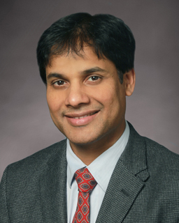
Sri-Rajasekhar (Raj) Kothapalli

Sri-Rajasekhar (Raj) Kothapalli

· Associate Professor of Biomedical EngineeringVerified

Pennsylvania State University · Acoustics

Active 2003–2026

h-index26
Citations2.5k
Papers11754 last 5y
Funding$1.8M1 active
See your match with Sri-Rajasekhar (Raj) Kothapalli — sign in to PhdFit.Sign in

About

Professor Sri-Rajasekhar (Raj) Kothapalli is the Principal Investigator of the BioPhotonics and Ultrasound Imaging Laboratory at Pennsylvania State University. He is an Associate Professor in the Department of Biomedical Engineering and also holds a courtesy appointment in the Department of Acoustics. Prof. Kothapalli received his foundational education and research training in Physics and Optical Engineering, culminating in a Ph.D. in Biomedical Engineering from Washington University in Saint Louis in 2009 under the mentorship of Prof. Lihong Wang. His doctoral research focused on the development of ultrasound-modulated optical tomography (UOT), where he hypothesized and demonstrated that UOT can image optical scattering contrast at ultrasonic resolution through theory, simulations, and experiments. He further explored various optical technologies such as CCD-based speckle contrast detection, Long cavity Fabry-Perot Interferometers, Four-wave mixing, and Spectral Hole Burning to efficiently detect ultrasound-modulated light photons. During his postdoctoral research at Stanford University from 2009 to 2013, Dr. Kothapalli designed, developed, and clinically translated a novel dual-modality Transrectal Ultrasound and Photoacoustic (TRUSPA) imaging device for human prostate cancer screening. This work was conducted under the guidance of Prof. Sanjiv Gambhir and Prof. Khuri-Yakub in the Departments of Radiology and Electrical Engineering, respectively. His notable contributions during this period include the first demonstration of Cerenkov luminescence endoscopy, photoacoustic molecular targeted imaging approaches using small molecules and nanoparticles, and the development of single cell photonic nanocavities for intracellular probing. From 2014 to 2016, he served as an instructor in the Department of Radiology at Stanford University, where he conducted first-in-human pilot clinical studies on prostate cancer patients using the TRUSPA device he developed. Prof. Kothapalli has been recognized with prestigious awards including the K99-R00 Pathway to Independence award from the National Institute of Biomedical Imaging and Bioengineering (NIBIB) of the NIH in 2014, and the NSF CAREER award from the Electrical, Communications, and Cyber Systems division of the National Science Foundation in 2023. Beyond his research and teaching activities, he enjoys gardening, playing squash, and music.

Research topics

  • Computer Science
  • Materials science
  • Physics
  • Optics
  • Acoustics
  • Optoelectronics
  • Composite material
  • Electrical engineering
  • Chemistry
  • Combinatorial chemistry
  • Biomedical engineering
  • Nanotechnology
  • Photochemistry
  • Electronic engineering
  • Telecommunications
  • Biochemistry
  • Mathematics
  • Organic chemistry
  • Biophysics

Selected publications

  • Integrated ultrasound stimulation and optical imaging of awake mice brain using a transparent ultrasound transducer cranial window

    npj Acoustics · 2026-01-08 · 3 citations

    articleSenior author
  • A novel optical elastography platform integrating transparent ultrasound transducer and digital holography

    2026-01-15

    articleSenior author
  • Portable laser diode-based multispectral photoacoustic and ultrasound imaging system for point-of-care screening applications

    2026-01-15

    articleSenior author
  • Multiparametric transrectal ultrasound and photoacoustic imaging system for prostate cancer diagnosis

    2026-03-04

    articleSenior author
  • Integrated Ultrasound Neuromodulation and Optical Neuroimaging in Awake Mice using a Transparent Ultrasound Transducer Cranial Window

    bioRxiv (Cold Spring Harbor Laboratory) · 2025-02-24

    preprintOpen accessSenior authorCorresponding

    Abstract Ultrasound neuromodulation is a rapidly advancing, non-invasive technique with significant therapeutic potential for treating various neurological disorders. Although extensive in vitro and in vivo studies have provided valuable insights into its modulatory effects, the underlying mechanisms remain poorly understood, limiting its clinical translation. Optical neuroimaging techniques can help investigate these mechanisms; however, the opacity and bulkiness of conventional ultrasound transducers pose significant challenges for their integration with in vivo ultrasound neuromodulation studies, particularly in awake rodents. To address these limitations, we propose a straightforward solution: a miniaturized lithium niobate-based transparent ultrasound transducer (TUT) integrated as a thinned-skull cranial window for ultrasound stimulation while facilitating multimodal optical neuroimaging in awake mice brain. Using laser speckle contrast imaging and intrinsic optical signal imaging, we studied changes in brain hemodynamics in response to various ultrasound stimulation sequences. Our experiments demonstrated that TUT cranial window can robustly induce neuromodulatory effects with observed increase in both cerebral blood flow and total hemoglobin, with peak and cumulative hemodynamic changes directionally correlated with ultrasound stimulation duration and intensity. Overall, these findings highlight that TUT cranial window can seamlessly integrate ultrasound stimulation and optical neuroimaging in awake mouse brain models, offering promising prospects for uncovering the underlying mechanisms of ultrasound neuromodulation.

  • Differentiating Aggressive from Nonaggressive Prostate Cancer Using Unconjugated Contrast Agents

    ACS Applied Materials & Interfaces · 2025-06-04 · 1 citations

    articleSenior authorCorresponding

    Prostate cancer is one of the most common malignancies among men globally. Early and accurate assessment of tumor aggressiveness is essential for guiding treatment decisions and improving patient outcomes. With growing clinical interest in combining optical, ultrasound, and photoacoustic imaging approaches for cancer detection, we systematically investigated the cellular uptake of three multimodal contrast agents─indocyanine green (ICG), ICG-loaded nanobubbles (ICG-NBs), and ICG-loaded microbubbles (ICG-MBs)─across prostate cancer cell lines with varying aggressiveness (PC-3M > PC-3 > DU-145 > LNCaP). Concentration- and time-dependent assays revealed that after 1 h of incubation, ICG-NBs exhibited 5-fold higher uptake (***p < 0.001) by highly metastatic PC-3M cells compared to the less aggressive and indolent cell lines. In contrast, ICG-MBs showed minimal uptake, detectable only after prolonged incubation (12 h) in PC-3M and PC-3 cells, while free ICG exhibited negligible uptake at 6 h, except in PC-3M cells, but increased over time in all cell lines. Validation across breast, colorectal, and pancreatic cancer cells further confirmed that ICG-NB uptake positively correlates with the metastatic potential of the cancer cell. Mechanistic studies identified macropinocytosis as the primary pathway for ICG-NB internalization, with additional contributions from clathrin-mediated endocytosis. These findings highlight that unconjugated ICG-NBs can selectively differentiate aggressive from indolent cancer phenotypes, offering a promising multimodal contrast agent for cancer theranostics.

  • Atherosclerosis-induced vascular contributions to cognitive impairment

    Physiology · 2025-05-01

    article

    Epidemiologic and pathological studies have indicated an association between vascular diseases and cognitive dysfunction, implicating vascular contributions to cognitive impairment and dementia (VCID). Atherosclerosis is a leading cause of vascular disease worldwide. Yet, the direct impact of atherosclerosis on cognitive impairment and related mechanisms remains largely unknown. Using an atherosclerosis mouse model, high fat diet (HFD)-fed ApoE -/- mice, our study found that atherosclerotic plaques not only formed in aorta and peripheral arteries, but also extensively developed in carotid and intracranial arteries. These observations led to our hypothesis that atherosclerotic blockages of extra/intra-cranial arteries cause cerebral hypoperfusion, vascular inflammation and remodeling, resulting in vascular pathology-induced cognitive impairment. ApoE -/- mice at 8 weeks of age were started on either normal or HFD (40% fat calories). The total cholesterol levels of HFD ApoE -/- mice exceeded 300 mg/dL within 1–2 weeks. By 16 weeks of HFD, extensive atherosclerotic blockages were detected in carotid and downstream arteries. At bifurcation regions, the blockages were 60–70% of the vessel lumen. Longitudinal behavior studies were performed before and after the start of HFD at 4-week intervals utilizing the novel object recognition (NOR) and Y-Maze memory tasks. Significant cognitive impairment was detected in ApoE -/- mice as early as 12 weeks of HFD and the levels of cognitive deficits were progressed with the time of HFD feeding. In contrast, age-matched control diet-fed ApoE -/- mice maintained proper memory performance for both tasks up to 32 weeks. We also performed in vivo ultrasound localization microscopy (ULM) in intact mouse brains. ULM images showed a reduction of both functional cerebral vascular density (21-35%) and blood volume (45-62%) in 4 brain vascular regions in the HFD mouse when compared with age-matched control. A high-fidelity image-based computational fluid dynamic (CFD) model was developed to predict atherosclerosis-induced hemodynamic alterations in cerebral vascular networks. A 65% lumen blockage of internal carotid artery predicts over 60% of flow reduction in major cerebral arteries. Using 3D whole mouse brain vascular images derived by light sheet fluorescence microscopy, CFD predicts a significantly increased spatial heterogeneity of the flow distribution in the HFD mouse versus the control, which predicted flow reductions in 75% of the major arteries and flow increases in 25% of the downstream vessels. In summary, both our in vivo animal model studies and in silico CFD predictions support our hypothesis, demonstrating atherosclerotic plaque deposition-induced cerebral hypoperfusion and vascular remodeling, which could be one of the key mechanisms for atherosclerosis-induced VCID. National Institutes of Health HL144620 and NINDS R01NS139128 This abstract was presented at the American Physiology Summit 2025 and is only available in HTML format. There is no downloadable file or PDF version. The Physiology editorial board was not involved in the peer review process.

  • Metabotissugenic citrate biomaterials orchestrate bone regeneration via citrate-mediated signaling pathways

    Science Advances · 2025-07-23 · 10 citations

    articleOpen access

    Bone regeneration requires coordinated anabolic and catabolic signaling, yet the interplay between mammalian target of rapamycin complex 1 (mTORC1) and adenosine monophosphate-activated protein kinase (AMPK) pathways remains unclear. This study reveals that citrate, glutamine, and magnesium synergistically activate both pathways via calcium/calmodulin-dependent protein kinase kinase 2 (CaMKK2)- and protein kinase B (Akt)-dependent signaling, bypassing the traditional adenosine monophosphate (AMP)/adenosine triphosphate (ATP) sensing mechanism. This dual activation supports sustained energy metabolism during osteogenesis and challenges the canonical antagonism between mTORC1 and AMPK. We developed CitraBoneQMg, a citrate-based biomaterial incorporating these components via one-pot synthesis. CitraBoneQMg provides sustained release, photoluminescent and photoacoustic imaging capabilities, and tunable mechanical properties. In vitro, it promotes osteogenesis by enhancing alkaline phosphatase (ALP) activity, osteogenic gene expression, and calcium deposition. In vivo, it accelerates bone regeneration in a rat calvarial defect model while promoting anti-inflammatory and neuroregenerative responses. We define this integrated effect as "metabotissugenesis," offering a metabolically optimized approach to orthopedic biomaterial design.

  • Multiwavelength laser diode based portable photoacoustic and ultrasound imaging system for point of care applications

    Journal of Biophotonics · 2024-05-02 · 10 citations

    articleOpen accessSenior authorCorresponding

    Vascular diseases are a leading cause of death and disability worldwide. Despite having precursor conditions like peripheral arterial disease (PAD), they are often only diagnosed after the onset of stroke or heart attack. Low-cost, portable, noninvasive, point-of-care (POC), label-free assessment of deep vascular function benefits PAD diagnosis, especially in resource poor settings of the world. Doppler ultrasound-based blood flow measurements can diagnose PAD, albeit with limited sensitivity and specificity. To overcome this, here, we propose the first-of-its-kind dual-modality photoacoustic-and-ultrasound (PAUS) imaging system that integrates a multiwavelength pulsed laser diode (PLD) with a compact ultrasound data acquisition unit. The mesoscopic imaging depth of the portable PLD-PAUS system was validated using tissue phantoms, and its multispectral photoacoustic imaging capabilities were validated using an atherosclerosis-mimicking phantom. Furthermore, we demonstrated high-contrast volumetric in vivo photoacoustic imaging of rodent abdominal vasculature and quantified vessel reactivity due to hypercapnia stimulation. The multiparametric functional and molecular imaging capabilities of the PLD-PAUS system holds promise for POC applications.

  • Multiparametric Brain Hemodynamics Imaging Using a Combined Ultrafast Ultrasound and Photoacoustic System

    Advanced Science · 2024-06-17 · 18 citations

    articleOpen accessSenior authorCorresponding

    Abstract Studying brain‐wide hemodynamic responses to different stimuli at high spatiotemporal resolutions can help gain new insights into the mechanisms of neuro‐ diseases and ‐disorders. Nonetheless, this task is challenging, primarily due to the complexity of neurovascular coupling, which encompasses interdependent hemodynamic parameters including cerebral blood volume (CBV), cerebral blood flow (CBF), and cerebral oxygen saturation (SO 2 ). The current brain imaging technologies exhibit inherent limitations in resolution, sensitivity, and imaging depth, restricting their capacity to comprehensively capture the intricacies of cerebral functions. To address this, a multimodal functional ultrasound and photoacoustic (fUSPA) imaging platform is reported, which integrates ultrafast ultrasound and multispectral photoacoustic imaging methods in a compact head‐mountable device, to quantitatively map individual dynamics of CBV, CBF, and SO 2 as well as contrast agent enhanced brain imaging at high spatiotemporal resolutions. Following systematic characterization, the fUSPA system is applied to study brain‐wide cerebrovascular reactivity (CVR) at single‐vessel resolution via relative changes in CBV, CBF, and SO 2 in response to hypercapnia stimulation. These results show that cortical veins and arteries exhibit differences in CVR in the stimulated state and consistent anti‐correlation in CBV oscillations during the resting state, demonstrating the multiparametric fUSPA system's unique capabilities in investigating complex mechanisms of brain functions.

Recent grants

Frequent coauthors

  • Sumit Agrawal

    Pennsylvania State University

    45 shared
  • Ajay Dangi

    Pennsylvania State University

    30 shared
  • Haoyang Chen

    Hebei Medical University

    30 shared
  • Chandra Yelleswarapu

    University of Massachusetts Boston

    22 shared
  • Shubham Mirg

    Pennsylvania State University

    16 shared
  • D. V. G. L. N. Rao

    Boston University

    15 shared
  • Pengfei Wu

    14 shared
  • Sanjiv S. Gambhir

    Stanford University

    13 shared

Labs

Education

  • Ph.D., Acoustics

    Pennsylvania State University

    1990
  • M.S., Acoustics

    University of Texas at Austin

    1986
  • B.S., Acoustics

    Indian Institute of Technology Madras

    1984

Awards & honors

  • NSF CAREER, National Science Foundation, March 2023 - Februa…
  • Best Paper Award, In Vivo Ultrasound Imaging GRC conference,…
  • Best Paper Award Runner-Up, IEEE Sensors Letters, November 2…
  • NIH Pathway to Independence Grant Award K99/R00, National In…
  • Resume-aware match score
  • Save to shortlist
  • AI-drafted outreach

See your match with Sri-Rajasekhar (Raj) Kothapalli

PhdFit ranks faculty by your research interests, methods, and publications — grounded in their actual work, not templates.

  • Free to start
  • No credit card
  • 30-second signup