Resume-aware faculty matching

Find professors who actually fit you

Upload your resume. Four AI agents analyze your background, rank the faculty who fit, inspect their recent research, and help you draft outreach — grounded in their actual work, not templates.

Free to startNo credit cardCancel anytime
Top matches Balanced preset
Dr. Sarah Chen
Stanford · Interpretability · NLP
91
Dr. Marcus Holloway
MIT · Robotics · RL
84
Dr. Aisha Okonkwo
CMU · Fairness · HCI
82
Nova · Professor Researcher · re-ranking top 20…
R. Joshua Dym

R. Joshua Dym

· Head of Emergency Radiology, Co-director of Quality Assurance, ProfessorVerified

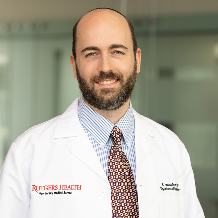
Rutgers University · Radiology

Active 2006–2024

h-index20
Citations1.3k
Papers5311 last 5y
Funding
See your match with R. Joshua Dym — sign in to PhdFit.Sign in

About

R. Joshua Dym, MD is a Professor of Radiology at Rutgers New Jersey Medical School and serves as the Section Head of Emergency Radiology. His areas of special interest include emergency abdominal imaging, emergency thoracic imaging, emergency neuroradiology, and radiology quality and safety. Dr. Dym has been widely published on these topics in prestigious imaging journals and several textbooks. He has a particular passion for radiology education, having co-authored four seminal educational review articles in Radiographics, which have been downloaded over 420,800 times as of July 2022. He has presented 36 educational exhibits at national meetings and delivered numerous educational talks regionally and nationally. Dr. Dym is a Senior Editor for the Academy of Online Radiology Education, where he edits an emergency radiology blog reviewing current research. In recognition of his contributions to radiology education, he received the 2020 RSNA Honored Educator Award, an honor awarded to only 49 radiologists nationwide that year. He has also received multiple awards for educational exhibits and research presentations at national meetings. Additionally, Dr. Dym serves as a reviewer for several journals and is active in national radiology societies, including the American Society of Emergency Radiology, where he is a member of the Mass Casualty Incidents Expert Panel. He holds a medical degree from Albert Einstein College of Medicine and a bachelor's degree from Brooklyn College of the City University of New York, and he is licensed to practice medicine in New Jersey.

Research topics

  • Medicine
  • Internal medicine
  • Radiology
  • Surgery

Selected publications

  • CPI Emergency & Trauma Imaging Module 2024

    2024-10-03

    dataset
  • “Learning From Disaster: What Past Events Can Teach Radiology Departments about Planning for a Mass Casualty Incident”

    Current Problems in Diagnostic Radiology · 2023-05-11 · 2 citations

    reviewOpen accessSenior authorCorresponding

    The increased frequency of mass shootings, terror attacks, and natural disasters in recent years have presented challenges to provision of quality medical care in both short and long-term stressful situations. While emergency departments and trauma surgeons are usually the face of the response to mass casualty incidents (MCI), other departments such as radiology are often active participants in caring for these patients but may not be as well prepared. In this article, we review nine papers describing the experiences of various radiology departments with specific MCIs and the lessons they learned from those experiences. By analysis of common themes raised in these papers, we hope to enable departments to incorporate these lessons into their disaster plans to enhance their preparedness for such events.

  • Can Gadolinium Be Used As An Alternative To Iodinated Contrast For CT Angiography During The Current Pandemic-Related Contrast Shortage?

    Journal of Medical Case Reports and Case Series · 2022-08-08 · 1 citations

    articleOpen access

    This halted the production of a water-soluble iodine contrast at a significant General Electric factory (of the GE Healthcare subsidiary).

  • Update on establishing and managing an overnight emergency radiology division

    Emergency Radiology · 2021-04-21 · 11 citations

    reviewOpen accessSenior author
  • Night and Day: Confounding Factors Complicate Comparison and Generalizability of Radiology Error Rates

    Radiology · 2020-12-15 · 2 citations

    letter1st authorCorresponding
  • Re: performing MRI on patients with MRI-conditional and non-conditional cardiac implantable electronic devices: an update for radiologists. A reply

    Clinical Radiology · 2020-03-12 · 1 citations

    letter
  • No way out: Causes of duodenal and gastric outlet obstruction

    Clinical Imaging · 2020 · 15 citations

    • Medicine
    • Radiology
    • Surgery
  • Performing MRI on patients with MRI-conditional and non-conditional cardiac implantable electronic devices: an update for radiologists

    Clinical Radiology · 2019-08-16 · 17 citations

    review
  • CT of the Neck: Image Analysis and Reporting in the Emergency Setting

    Radiographics · 2019-10-01 · 29 citations

    review

    Interpreting findings seen at CT of the neck is challenging owing to the complex and nuanced anatomy of the neck, which contains multiple organ systems in a relatively small area. In the emergency department setting, CT is performed to investigate acute infectious or inflammatory symptoms and chronic processes. With few exceptions, neck CT should be performed with intravenous contrast material, which accentuates abnormally enhancing phlegmonous and neoplastic tissues and can be used to delineate any abscesses or necrotic areas. As part of the evaluation, the vascular structures and aerodigestive tract must be scrutinized, particularly for patency. Furthermore, although the patient may present because of symptoms that suggest non–life-threatening conditions involving structures such as the teeth or salivary glands, there may be serious implications for other areas, such as the orbits, brain, and spinal cord, that also may be revealed at the examination. With a focus on the emergency setting, the authors propose using an approach to interpreting neck CT findings whereby 12 areas are systematically evaluated and reported on: the cutaneous and subcutaneous soft tissues, aerodigestive tract and adjacent soft tissues, teeth and periodontal tissues, thyroid gland, salivary glands, lymph nodes, vascular structures, bony airspaces, cervical spine, orbits and imaged brain, lung apices, and superior mediastinum. The use of a systematic approach to interpreting neck CT findings is essential for identifying all salient findings, recognizing and synthesizing the implications of these findings to formulate the correct diagnosis, and reporting the findings and impressions in a complete, clear, and logical manner. Online supplemental material is available for this article. ©RSNA, 2019

  • Implantable Electronic Stimulation Devices from Head to Sacrum: Imaging Features and Functions

    Radiographics · 2019-07-01 · 35 citations

    review

    Electronic stimulation devices are implanted in various locations in the body to decrease pain, modulate nerve function, or stimulate various end organs. The authors describe these devices using a craniocaudal approach, first describing deep brain stimulation (DBS) devices and ending with sacral nerve stimulation (SNS) devices. The radiology-relevant background information for each device and its imaging appearance are also described. These devices have a common design theme and include the following components: (a) a pulse generator that houses the battery and control electronics, (b) an insulated lead or wire that conveys signals to the last component, which is (c) an electrode that contacts the end organ and senses and/or acts on the end organ. DBS electrodes are inserted into various deep gray nuclei, most commonly to treat the symptoms of movement disorders. Occipital, trigeminal, and spinal nerve stimulation devices are used as second-line therapy to control craniofacial or back pain. For cardiac devices, the authors describe two newer devices, the subcutaneous implantable cardioverter defibrillator and the leadless pacemaker, both of which avoid complications related to having leads threaded through the venous system. Diaphragmatic stimulation devices stimulate the phrenic nerve to restore diaphragmatic movement. Gastric electrical stimulation devices act on various parts of the stomach for the treatment of gastroparesis or obesity. Finally, SNS devices are used to modulate urinary and defecatory functions. Common complications diagnosed at imaging include infection, hematoma, lead migration, and lead breakage. Understanding the components, normal function, and normal imaging appearance of each device allows the radiologist to identify complications. ©RSNA, 2019

Frequent coauthors

  • Meir H. Scheinfeld

    The Bronx Defenders

    64 shared
  • Alain Cunqueiro

    Albert Einstein College of Medicine

    38 shared
  • Peter Lee

    25 shared
  • William A. Gomes

    25 shared
  • Michael L. Lipton

    Albert Einstein College of Medicine

    17 shared
  • Benjamin H. Taragin

    University of Pennsylvania

    13 shared
  • Jonathan Sterman

    Montefiore Medical Center

    13 shared
  • Michael Spektor

    12 shared

Education

  • M.D.

    Albert Einstein College of Medicine

    2005
  • B.S.

    Brooklyn College of the City University of New York

    2001

Awards & honors

  • 2020 RSNA Honored Educator Award
  • Certificate of Achievement from the Academy of Radiology Lea…
  • Distinguished Reviewer for Abdominal Radiology
  • Resume-aware match score
  • Save to shortlist
  • AI-drafted outreach

See your match with R. Joshua Dym

PhdFit ranks faculty by your research interests, methods, and publications — grounded in their actual work, not templates.

  • Free to start
  • No credit card
  • 30-second signup