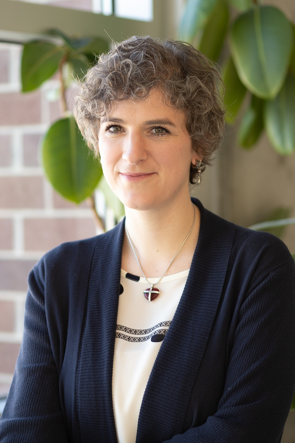
Julia Brasch
· Assistant ProfessorVerifiedUniversity of Utah · Biology
Active 1969–2025
About
Julia Brasch is an Assistant Professor of Biochemistry at the University of Utah, with a background that includes a B.S. and M.S. from Gottfried Wilhelm Leibniz University of Hannover, where she also earned her Ph.D. Her research focuses on understanding the molecular architecture of synapses in the brain, specifically how neurons form complex neural circuits through specialized junctions called synapses. Her laboratory aims to elucidate the protein complexes involved in synaptic formation, establishment, and properties, with a particular emphasis on synaptic cell adhesion molecules. Her work employs advanced techniques such as cryo-electron microscopy, electron cryo-tomography, and biophysical methods to analyze protein interactions at a molecular level. The goal of her research is to identify the building blocks of the synapse and understand how these elements are assembled, which contributes to a broader understanding of the nervous system's extracellular architecture. Her contributions include detailed structural and biophysical analysis of cell adhesion molecules and their interactions, advancing knowledge in synaptic biology and molecular neuroscience.
Research topics
- Biophysics
- Biology
- Cell biology
- Biochemistry
- Chemistry
- Computational biology
- Evolutionary biology
Selected publications
T cell ectosomes promote antibody responses through cognate TCR-pMHC interactions
bioRxiv (Cold Spring Harbor Laboratory) · 2025-08-02
preprintOpen accessProtective antibody-mediated immunity requires effective T cell-mediated help. Recognition of peptide antigens presented by major histocompatibility complex class II molecules (pMHCII), via cognate T cell antigen receptors (TCR), activates CD4+ T helper cells to upregulate expression of CD40L and helper cytokines. CD40L-CD40 interactions at T-B cell synapses and secreted cytokines are well established mediators of T cell help. Whether engaged pMHCII also transmits signals that help B cells remains unresolved. Here, we show that TCR-enriched nanoscale vesicles shed by activated T cells (ectosomes) are transferred to antigen-primed B cells, where they engage and cluster cognate pMHCII, triggering signaling and specific IgG antibody production. Disruption of ectosome release attenuates B cell antibody production, while native and synthetic ectosomes boost antibody responses. We conclude that T cell ectosomes constitute a new modality of help for B cells, delivered through engagement of pMHCII by cognate ectosomal TCR.
Progress in Neurobiology · 2025-05-01 · 3 citations
articleCryoEM/ET 101: An Engaging Online Self-Paced Course That Teaches Cryo-EM and Cryo-ET Principles
Microscopy and Microanalysis · 2024-07-01
articleOpen accessSenior authorCryo-electron microscopy and tomography (cryo-EM and -ET) have emerged as indispensable tools to visualize high-resolution details of biological structures across a broad range of scales, from small molecules and proteins to cells and tissues.Although there is growing enthusiasm for these methods, there are barriers to starting.First, these methods require expensive equipment; second, there is a steep learning curve for newcomers to the field, especially since there are not experts available at every institution; third, there are only inadequate and dispersed learning materials for beginners.Access to equipment and expertise has been made available through the establishment of national cryo-EM and cryo-ET centers [1], which accept trainees of all experience levels and has already boosted the use of the method.To address the gap in training and the remaining limitations, we have designed a media-rich and user-friendly approach in the form of a self-paced, interactive online course that emphasizes practical training for cryo-EM and cryo-ET novices [2].Our freely available course engages and teaches newcomers the basics of the typical cryo-EM and cryo-ET project workflows, which includes best practices in sample and grid preparation, data collection, and data processing.The highlights are animations that make complex concepts easily digestible, interactives to solidify understanding and practical videos of commonly used methods such as plunge freezing, sample insertion into instruments, and data collection.Image galleries of real examples showcase 'the good, the bad, and the ugly', which provide real world outcomes of TEM imaging.Our course materials have been implemented by the NIH National Cryo-EM and Cryo-ET centers to support the teaching of cryo-EM/ET methods to newcomers and to augment their preparation for on-site visits.CryoEM-101 accelerates learning time and helps center staff to be more efficient by allowing them to focus on hands-on training rather than teaching basic concepts.Users can then take the methods back to their home institutions.We found that users access the materials for their own learning, for teaching in a classroom setting, as trainees newly joining cryo-EM/ET focused labs, and more.Furthermore, the course materials are used in the National Centers' Merit Badge program, which certifies users of certain skills across national centers.Overall, the goal of our course is to guide cryo-EM and cryo-ET newcomers to a point along their training where they can independently prepare and optimize their specimens for high-quality data collection at dedicated EM centers.This resource has established a broad user base globally [3].
Abstract 1962: Structure and mechanism of Neurexin - Adhesion GPCR interactions in neuronal Synapses
Journal of Biological Chemistry · 2023-01-01
articleOpen accessSenior authorNucleic Acids Research · 2023-01-30 · 23 citations
articleOpen accessThe conserved complex of the Rad6 E2 ubiquitin-conjugating enzyme and the Bre1 E3 ubiquitin ligase catalyzes histone H2B monoubiquitination (H2Bub1), which regulates chromatin dynamics during transcription and other nuclear processes. Here, we report a crystal structure of Rad6 and the non-RING domain N-terminal region of Bre1, which shows an asymmetric homodimer of Bre1 contacting a conserved loop on the Rad6 'backside'. This contact is distant from the Rad6 catalytic site and is the location of mutations that impair telomeric silencing in yeast. Mutational analyses validated the importance of this contact for the Rad6-Bre1 interaction, chromatin-binding dynamics, H2Bub1 formation and gene expression. Moreover, the non-RING N-terminal region of Bre1 is sufficient to confer nucleosome binding ability to Rad6 in vitro. Interestingly, Rad6 P43L protein, an interaction interface mutant and equivalent to a cancer mutation in the human homolog, bound Bre1 5-fold more tightly than native Rad6 in vitro, but showed reduced chromatin association of Bre1 and reduced levels of H2Bub1 in vivo. These surprising observations imply conformational transitions of the Rad6-Bre1 complex during its chromatin-associated functional cycle, and reveal the differential effects of specific disease-relevant mutations on the chromatin-bound and unbound states. Overall, our study provides structural insights into Rad6-Bre1 interaction through a novel interface that is important for their biochemical and biological responses.
Structures of LRP2 reveal a molecular machine for endocytosis
Cell · 2023 · 69 citations
- Biology
- Cell biology
- Biochemistry
Visualizing cadherin intermembrane adhesion assemblies using cryo-electron tomography
Microscopy and Microanalysis · 2021-07-30
articleOpen accessSenior authorCadherins are a superfamily of transmembrane proteins that contain extracellular cadherin (EC) domains, barrels connected by linker regions that typically bind calcium ions adding rigidity to their structure (1). Classical cadherins, the most well-characterized sub-branch of the superfamily, are adhesion molecules that form intracellular adherens junctions that are key to the development and maintenance of tissue architecture from epithelia (2) to the central nervous system (3). Classical cadherins mediate adhesion by trans interaction with a cognate cadherin molecule on an opposing membrane surface, the interface of which involves a conserved strand swapping mechanism between the membrane distal EC1 domains (4-6). The two subfamilies of vertebrate classical cadherinstype I and type IIboth contain five EC domains, a single-pass transmembrane region and a cytoplasmic domain that interacts with the actin cytoskeleton through -and catenin, but vary in their expression patterns and the exact details of the EC1 strand-swapping interaction. Type I cadherins, like other adhesion molecules ( As expected, mutations in type I cadherins that disrupt trans interactions were found to completely ablate all adhesion, while mutations targeting the cis interface prevent the formation of ordered assemblies in crystal structures, on liposomes and on the cellular level (5). However, whether type II cadherins form ordered assemblies is as yet unknown. Structural characterization of type II cadherins is an important step towards understanding their potential role in downstream signaling or the conveyance of force between opposing cells.
Computational model of E-cadherin clustering under force
Biophysical Journal · 2021 · 17 citations
- Cell biology
- Biophysics
- Chemistry
Crystal structure of human protocadherin 8 EC5-EC6
2020-01-06
paratextCrystal structure of monomeric human protocadherin 10 EC1-EC4
2020-01-06
paratext
Frequent coauthors
- 43 shared
Barry Honig
Columbia University
- 37 shared
Lawrence Shapiro
Columbia University
- 29 shared
O.J. Harrison
National Institute of Allergy and Infectious Diseases
- 24 shared
Göran Ahlsén
Columbia University
- 22 shared
Phinikoula S. Katsamba
Columbia University
- 20 shared
Priyamvada Acharya
Duke University
- 19 shared
Bridget Carragher
Chan Zuckerberg Initiative (United States)
- 19 shared
Fabiana Bahna
Columbia University
Labs
Education
B.S.
Gottfried Wilhelm Leibniz University of Hannover
M.S.
Gottfried Wilhelm Leibniz University of Hannover
Ph.D.
Gottfried Wilhelm Leibniz University of Hannover
- Resume-aware match score
- Save to shortlist
- AI-drafted outreach
See your match with Julia Brasch
PhdFit ranks faculty by your research interests, methods, and publications — grounded in their actual work, not templates.
- Free to start
- No credit card
- 30-second signup