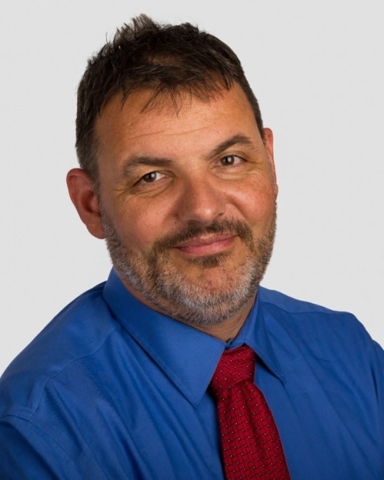
Urs Utzinger
· Associate Professor of Biomedical Engineering Associate Professor of Obstetrics and Gynecology Associate Professor of Electrical and Computer Engineering Associate Professor, BIO5 Institute Associate Professor of Optical Sciences Associate Department Head for Assessment and Accreditation Member of the Graduate FacultyVerifiedUniversity of Arizona · Biomedical Engineering
Active 1992–2025
About
Urs Utzinger is an associate professor of biomedical engineering with additional appointments in obstetrics and gynecology, optical sciences, electrical and computer engineering, and the BIO5 Institute at the University of Arizona. He received his engineering degree (Dipl. Ing. ETH) in mechanical engineering in 1989 and his PhD (Dr. sc. techn. ETH) in biomedical engineering in 1995 from the Swiss Federal Institute of Technology in Zürich, Switzerland. His post-doctoral work was conducted at The University of Texas, Austin, in the Optical Spectroscopy Laboratory of Rebecca Richards-Kortum. Utzinger's research focuses on developing clinical imaging instrumentation to evaluate gynecological and gastrointestinal cancers. He has studied the extracellular matrix and angiogenesis using microscopy techniques and has developed spectral imaging techniques for skin. He has also contributed to the design of hands-on courses for biomedical engineering students, including a sophomore design course and a medical device design course. In 2016, he developed the Salter Medical Device Laboratory, which he designed himself. Throughout his career, Utzinger has been involved in advancing optical imaging technologies, including optical coherence tomography, multispectral fluorescence imaging, and the use of FDA-approved drugs as fluorescent dyes for optical biopsies. His work aims to improve early detection and diagnosis of cancers, particularly ovarian and colorectal cancers, through innovative imaging methods. He has served as interim department head in biomedical engineering and currently serves as associate department head of assessment and accreditation, contributing to the growth and accreditation of the program.
Research topics
- Computer Science
- Optics
- Medicine
- Engineering
- Psychology
- Mathematical analysis
- Chemistry
- Materials science
- Mathematics
- Pathology
- Biomedical engineering
- Biochemistry
- Computational physics
- Physics
- Biology
- Engineering management
- Biotechnology
- Mathematics education
- Pedagogy
Selected publications
Proceedings on CD-ROM - International Society for Magnetic Resonance in Medicine. Scientific Meeting and Exhibition/Proceedings of the International Society for Magnetic Resonance in Medicine, Scientific Meeting and Exhibition · 2025-09-16
articleMotivation: Focused ultrasound (FUS) with microbubbles is a promising technology for opening the blood-brain barrier (BBB) to deliver therapeutics to the brain. Answering fundamental questions about the process of BBB opening will aid in the translation of this method to the clinic. Goal(s): Use FUS, 2-photon microscopy, MRI and immunohistochemistry to investigate FUS-induced changes in wild type and 5X-FAD AD mice. Approach: A cross-sectional study involving MRI, 2PM, and IHC to examine BBB opening at multiple timepoints in a disease model. Results: Results suggest BBB opening occurs in capillaries and that the BBB opening is similar between WT and 5X-FAD mice. Impact: DCE imaging and sub-micron imaging of the microvasculature after BBB opening provide insight into which vessels are opened after FUS and improve pharmacokinetic understanding of the paravascular space in an Alzheimer's Disease model.
Lasers in Surgery and Medicine · 2024-12-19 · 1 citations
articleOBJECTIVES: To study the healing response of rat Achilles tendon when lacerated or treated with intense therapeutic ultrasound (ITU) via utilization of multiphoton microscopy (MPM) imaging and histology. MATERIALS AND METHODS: The right Achilles tendon of each Sprague Dawley rat within a cohort was partially lacerated. 1 to 2 days post-surgery, each rat received ITU treatment of the Achilles tendon on either the right or left leg. Rats were euthanized in groups at 1, 3, 7, 14, or 28 days posttreatment and their tendons were explanted, formalin fixed, paraffin embedded, sectioned, and placed on slides for imaging. Slides from each time point were imaged using a laboratory built MPM with a 780 nm Ti:Sapphire laser. The resulting second harmonic generation (SHG) and two-photon excited fluorescence (2PEF) signals were captured, assessed, and compared to brightfield microscopy images of the same section subsequently stained with hematoxylin and eosin. RESULTS: At early timepoints, 2PEF images show the presence of red blood cells, infiltration of inflammatory cells and formation of a fibrin clot at laceration sites, and attraction of fibroblasts to ITU coagula. SHG images indicate an absence of organized collagen in both types of lesions. At later timepoints, new organized collagen can be seen at the laceration sites, and the concentration of inflammatory cells has noticeably decreased. Automated detection of red blood cells and infiltrative cells, as well as analysis of SHG signal intensity and homogeneity was performed at laceration locations. Results show that all quantities except SHG signal intensity approach normal values by day 28. Thus, combined analysis of 2PEF and SHG images elucidates tendon healing processes that align with and complement histological findings. CONCLUSION: These results indicate that multiphoton imaging can effectively visualize the healing response to mechanical (laceration) and thermal (ITU) injury, including the organization of new collagen which is more difficult to visualize with histology.
Proceedings on CD-ROM - International Society for Magnetic Resonance in Medicine. Scientific Meeting and Exhibition/Proceedings of the International Society for Magnetic Resonance in Medicine, Scientific Meeting and Exhibition · 2024-11-26
articleMotivation: The gold-standard for confirmation of blood-brain barrier (BBB) disruption after FUS is T1-weighted MRI with contrast enhancement. Conventional MRI-based metrics cannot differentiate between BBB leakage at the microvascular level, and thus there is a need for validation with higher spatial and temporal resolution modalitiesGoal(s): Use 2-photon microscopy to investigate solute extravasation from the microvasculature into the parenchyma and paravascular space. Approach: This project is an in-vivo validation of BBB opening after FUS using 2-photon microscopy and MRI. Results: Results suggest BBB opening occurs in capillaries, and that dye extravasation can be measured within minutes of sonication. Impact: Sub-micron imaging of the microvasculature after BBB opening will provide insight into which vessels are opened after FUS and improve pharmacokinetic understanding of the paravascular space around arterioles and venules.
A domed window chamber for multi-modality optical imaging
BioTechniques · 2024-11-25 · 1 citations
articleOpen accessmultimodality imaging with OCT, MPM, and LSFI was demonstrated.
Methods and Applications in Fluorescence · 2021 · 2 citations
- Pathology
- Biomedical engineering
- Medicine
staining of tissue, enabling real-time optical biopsies and other advanced microscopy technologies, which have implications for the speed and performance of tissue- and cellular-level diagnostics.
Biomedical Engineering Education · 2020 · 4 citations
- Computer Science
- Computer Science
- Engineering
Advances in Electrical and Computer Engineering · 2020 · 4 citations
- Optics
- Materials science
- Physics
A non-contact optical system has been designed to determine the absorption and reduced scattering coefficients of optical phantoms. The frequency-domain multi-distance method, which allows an estimation of optical properties in biological tissue uses the phase and intensity of radio frequency modulated light. The proposed design has been evaluated with optical phantoms. Estimated values for an absorption coefficient equal to 1 cm-1 are 0.795, 0.690, 0.670 and 0.613 cm-1 for wavelengths of 658 nm, 705 nm, 785 nm and 833 nm, respectively and for a reduced scattering coefficient equal to 22 cm-1, the estimated values are 19.876, 18.845, 17.134 and 17.927 cm-1. It has been concluded that this novel non-contact design can be used to determine the absorption and reduced scattering coefficients of optical phantoms. This system is the first step in medical equipment that may be used to measure absolute quantification of HbO, Hb, HbCO and HbMet concentrations in a contactless manner. Current oximeters with hemoglobin measurement capability require contact between the sensor and the skin. These oximeters have drawbacks when measuring child patients with asthma, bronchiolitis and bronchopneumonia. Currently it is not possible to assess oxygenation in open wounds. Therefore, it is worthwhile to develop a non-contact oximeter.
Racioethnic differences in the biomechanical response of the lamina cribrosa
Acta Biomaterialia · 2019-02-23 · 28 citations
articleBiomechanical Response of the Lamina Cribrosa in Glaucomatous and Non glaucomatous samples
Investigative Ophthalmology & Visual Science · 2019-07-22
articleOpen accessPerformance of combined OCT/MFI microendoscope for ovarian cancer detection
2018-01-25
articleTo improve early detection of ovarian cancer, we have designed and built a microendoscope that combines optical coherence tomography (OCT) and multispectral fluorescence imaging (MFI) into a 0.7 mm diameter package. An endoscope of this size allows access to the ovaries through the fallopian tubes creating a minimally invasive procedure. We characterize the falloposcope’s imaging behavior and show that this system provides contrast on ex vivo surgical samples of ovary and fallopian tube. In addition, we show the mechanical performance of the endoscope in an anatomically correct model of the female reproductive treatment.
Recent grants
NIH · $458k · 2009
NIH · $915k · 2008
Frequent coauthors
- 74 shared
Rebecca Richards‐Kortum
Rice University
- 43 shared
Jennifer K. Barton
- 32 shared
Molly Brewer
UConn Health
- 31 shared
Michele Follen Mitchell
- 28 shared
Carrie Brookner
- 28 shared
Michele Follen
- 24 shared
Jonathan P. Vande Geest
McGowan Institute for Regenerative Medicine
- 22 shared
Nathaniel D. Kirkpatrick
Education
- 1995
Dr. sc. Tech., Biomedical Engineering
Eidgenössische Technische Hochschule Zürich
- 1989
dipl. Ing. ETH, Biomedical Engineering
Eidgenössische Technische Hochschule Zürich
- Resume-aware match score
- Save to shortlist
- AI-drafted outreach
See your match with Urs Utzinger
PhdFit ranks faculty by your research interests, methods, and publications — grounded in their actual work, not templates.
- Free to start
- No credit card
- 30-second signup