Resume-aware faculty matching

Find professors who actually fit you

Upload your resume. Four AI agents analyze your background, rank the faculty who fit, inspect their recent research, and help you draft outreach — grounded in their actual work, not templates.

Free to startNo credit cardCancel anytime
Top matches Balanced preset
Dr. Sarah Chen
Stanford · Interpretability · NLP
91
Dr. Marcus Holloway
MIT · Robotics · RL
84
Dr. Aisha Okonkwo
CMU · Fairness · HCI
82
Nova · Professor Researcher · re-ranking top 20…
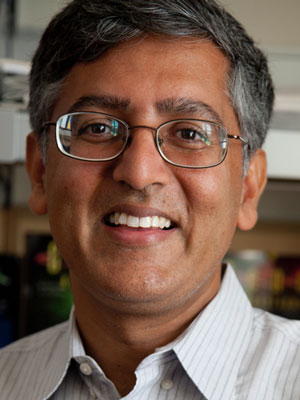
Arshad Desai

Arshad Desai

· Professor and ChairVerified

University of California, San Diego · Cell and Developmental Biology

Active 1995–2026

h-index101
Citations32.1k
Papers34358 last 5y
Funding$18.7M1 active
See your match with Arshad Desai — sign in to PhdFit.Sign in

About

Arshad Desai is a professor at the University of California, San Diego, involved in research within the Oegema and Desai Labs. His work focuses on understanding the mechanisms of cell division, particularly the regulation of spindle microtubule dynamics and attachments during mitosis. As a key member of the lab, he contributes to exploring how spindle poles regulate kinetochore–microtubule interactions to ensure accurate chromosome segregation and proper spindle size. His research employs model organisms such as C. elegans to elucidate fundamental cellular processes related to cell division, with implications for understanding developmental biology and disease mechanisms.

Research topics

  • Cell biology
  • Biology
  • Genetics
  • Chemistry
  • Physics
  • Biochemistry

Selected publications

  • Inhibitor-2 directs formation of PP1 holoenzymes through a docking motif-dependent transfer of catalytic subunits to adapters

    bioRxiv (Cold Spring Harbor Laboratory) · 2026-03-17

    articleOpen accessSenior authorCorresponding

    The catalytic subunits of protein phosphatase 1 (PP1) achieve spatiotemporal substrate specificity by assembling with diverse regulatory adapters to form holoenzymes. Three conserved proteins-Sds22, Inhibitor-2 and Inhibitor-3-facilitate loading of PP1 catalytic subunits (PP1cs) onto adapters. We show here that Inhibitor-2 is central to a dynamic cycle that directs formation of adapter-bound PP1 holoenzymes. Inhibitor-2 engages PP1cs via two adapter-like docking motifs (RVxF and SILK) and an active site-binding inhibitory region. While Inhibitor-2 depletion produced moderate phenotypes, mutation of its RVxF docking motif caused severe defects resembling global PP1c inhibition. The RVxF mutant did not prevent PP1c binding or reduce PP1c stability but inhibited formation of adapter-bound holoenzymes. The severe effects of the RVxF mutation were suppressed by linked mutation of the inhibitory active site-binding motif. These results suggest that Inhibitor-2 is integral to a dynamic cycle that delivers PP1cs to adapters, with its RVxF motif being critical for coupling relief of active site inhibition to adapter handoff.

  • The mitotic stopwatch synergizes with mild p53 activation to halt cell proliferation

    bioRxiv (Cold Spring Harbor Laboratory) · 2025-12-03

    articleOpen access

    ABSTRACT The mitotic stopwatch suppresses proliferation of cell lineages experiencing prolonged mitosis that are prone to chromosome missegregation and tumorigenesis. It converts extended mitotic duration into heritable USP28–53BP1 complexes that stabilize p53 and accumulate over generations. To identify genes whose knockout activates the stopwatch, we performed a CRISPR/Cas9 screen comparing dropout kinetics of essential-gene gRNAs in cells lacking versus possessing the stopwatch. Two classes of knockouts emerged: one (27/60 top hits) that prolonged mitosis, and another (33/60 top hits) that mildly elevated p53 without significant mitotic defects, indicating that the stopwatch synergizes with mild p53 activation to halt proliferation. Mild p53 elevation lowered the stopwatch complex threshold for daughter cell arrest and slightly prolonged mitosis. Integrated over successive divisions, the cumulative effect of multiple short mitotic extensions triggered stopwatch-dependent arrest. Thus, the mitotic stopwatch endows the p53 network with a durable lineage memory of modest stress, explaining its tumor-suppressive role.

  • Author Correction: A two-step mechanism for epigenetic specification of centromere identity and function

    Nature Cell Biology · 2025-06-23

    erratumOpen access
  • A chromatin-associated pool of Aurora A controls kinetochore-microtubule attachments to ensure chromosome biorientation

    bioRxiv (Cold Spring Harbor Laboratory) · 2025-11-01

    preprintOpen accessSenior authorCorresponding

    Abstract Accurate chromosome segregation requires dynamic kinetochore–microtubule attachments that, under the regulation of Aurora family kinases, biorient and align replicated chromosomes. In C. elegans , Aurora A acts with the TPX2-related activator TPXL-1 to regulate these attachments and control spindle length. We show that, in addition to prominent spindle pole localization, TPXL-1–AurA has a chromatin-associated pool positioned between the sister kinetochores. Structural modeling and biochemical analysis support TPXL-1 directly recognizing the nucleosome acidic patch via an arginine anchor. Disrupting this interaction selectively removed chromatin-bound TPXL-1–AurA and caused chromosome missegregation, whereas elevation of the chromatin pool disrupted chromosome alignment. These opposing perturbations inversely affected kinetochore recruitment of the microtubule-binding Ska complex. These results support spatially distinct TPXL-1–AurA populations acting sequentially, with the spindle pole pool controlling spindle length by switching kinetochores out of a depolymerization-coupled state, and the chromatin pool controlling attachment stabilization to ensure biorientation prior to anaphase.

  • Hybrid incompatibility emerges at the one-cell stage in interspecies Caenorhabditis embryos

    Current Biology · 2025-07-01 · 1 citations

    article
  • TRIM37 prevents ectopic spindle pole assembly by peptide motif recognition and substrate-dependent oligomerization

    Nature Structural & Molecular Biology · 2025-05-25 · 3 citations

    article
  • Phosphorylation remodels the mitotic centrosome matrix to generate bipartite γ-tubulin complex docking sites

    bioRxiv (Cold Spring Harbor Laboratory) · 2025-11-21

    preprintOpen access

    Abstract Mitotic centrosomes consist of centrioles surrounded by a proteinaceous matrix that docks and activates γ-tubulin complexes (γTuCs) to nucleate microtubules for spindle assembly. During mitotic entry, phosphorylation at centrosomes remodels CDK5RAP2 family matrix proteins to generate γTuC docking sites. We address the mechanism of this conversion using C. elegans SPD-5 as a model. We show that SPD-5 contains two regions, PRGB1 and PRGB2, that are each sufficient for Polo-Like Kinase 1 (PLK1) phosphorylation–regulated γTuC binding. We define key phosphosites in each region and uncover autoinhibition mediated by interactions within and between them. PRGB2 is dimeric and requires γTuCs containing the Mozart family microprotein MZT-1 for binding, whereas PRGB1 is monomeric and binds independently of MZT-1. Our results support PLK1 phosphorylation inducing a conformational change that enables MZT-1–dependent PRGB2 binding, which in turn relieves PRGB1 inhibition. Such a multi-step mechanism would ensure robust spindle assembly by restricting microtubule nucleation in space and time.

  • Phospho-KNL-1 recognition by a TPR domain targets the BUB-1–BUB-3 complex to <i>C. elegans</i> kinetochores

    bioRxiv (Cold Spring Harbor Laboratory) · 2024-02-11

    preprintOpen accessSenior authorCorresponding

    ABSTRACT During mitosis, the Bub1-Bub3 complex concentrates at kinetochores, the microtubule-coupling interfaces on chromosomes, where it contributes to spindle checkpoint activation, kinetochore-spindle microtubule interactions, and protection of centromeric cohesion. Bub1 has a conserved N-terminal tetratricopeptide (TPR) domain followed by a binding motif for its conserved interactor Bub3. The current model for Bub1-Bub3 localization to kinetochores is that Bub3, along with its bound motif from Bub1, recognizes phosphorylated “MELT” motifs in the kinetochore scaffold protein Knl1. Motivated by the greater phenotypic severity of BUB-1 versus BUB-3 loss in C. elegans , we show that the BUB-1 TPR domain directly recognizes a distinct class of phosphorylated motifs in KNL-1 and that this interaction is essential for BUB-1–BUB-3 localization and function. BUB-3 recognition of phospho-MELT motifs additively contributes to drive super-stoichiometric accumulation of BUB-1–BUB-3 on its KNL-1 scaffold during mitotic entry. Bub1’s TPR domain interacts with Knl1 in other species, suggesting that collaboration of TPR-dependent and Bub3-dependent interfaces in Bub1-Bub3 localization and functions may be conserved.

  • Kinetochore dynein is sufficient to biorient chromosomes and remodel the outer kinetochore

    Nature Communications · 2024-10-21

    articleOpen accessSenior author

    Multiple microtubule-directed activities concentrate on mitotic chromosomes to ensure their faithful segregation. These include couplers and dynamics regulators localized at the kinetochore, the microtubule interface built on centromeric chromatin, as well as motor proteins recruited to kinetochores and chromatin. Here, we describe an in vivo approach in the C. elegans one-cell embryo in which removal of the major microtubule-directed activities on mitotic chromosomes is compared to the selective presence of individual activities. Our approach reveals that the kinetochore dynein module, comprised of cytoplasmic dynein and its kinetochore-specific adapters, is sufficient to biorient chromosomes; by contrast, this module is unable to support congression. In coordination with orientation, the dynein module directs removal of outermost kinetochore components, including dynein itself, independently of the other microtubule-directed activities and kinetochore-localized protein phosphatase 1. These observations indicate that the kinetochore dynein module is sufficient to biorient chromosomes and to direct remodeling of the outer kinetochore in a microtubule attachment state-sensitive manner.

  • Control of cell proliferation by memories of mitosis

    Science · 2024-03-28 · 70 citations

    articleOpen accessSenior authorCorresponding

    Mitotic duration is tightly constrained, and extended mitosis is characteristic of problematic cells prone to chromosome missegregation and genomic instability. We show here that mitotic extension leads to the formation of p53-binding protein 1 (53BP1)–ubiquitin-specific protease 28 (USP28)–p53 protein complexes that are transmitted to, and stably retained by, daughter cells. Complexes assembled through a Polo-like kinase 1–dependent mechanism during extended mitosis and elicited a p53 response in G 1 that prevented the proliferation of the progeny of cells that experienced an approximately threefold extended mitosis or successive less extended mitoses. The ability to monitor mitotic extension was lost in p53-mutant cancers and some p53–wild-type (p53-WT) cancers, consistent with classification of TP53BP1 and USP28 as tumor suppressors. Cancers retaining the ability to monitor mitotic extension exhibited sensitivity to antimitotic agents.

Recent grants

Frequent coauthors

  • Karen Oegema

    University of California, San Diego

    318 shared
  • Pablo Lara-González

    97 shared
  • Paul S. Maddox

    University of North Carolina at Chapel Hill

    71 shared
  • Dhanya K. Cheerambathur

    Wellcome Centre for Cell Biology

    66 shared
  • Shaohe Wang

    Howard Hughes Medical Institute

    59 shared
  • Rebecca A. Green

    Bristol-Myers Squibb (United States)

    54 shared
  • Julien Dumont

    Université Paris Cité

    46 shared
  • Reto Gassmann

    i3S - Instituto de Investigação e Inovação em Saúde, Universidade do Porto

    45 shared

Labs

Education

  • PhD

    University of California San Francisco

    1997
  • Biology B.S.

    California State University East Bay

    1991

Awards & honors

  • Damon Runyon Scholar Award
  • American Society for Cell Biology Early Career Award
  • Keith Porter Fellowship
  • Lifetime Fellow of the American Society for Cell Biology
  • Resume-aware match score
  • Save to shortlist
  • AI-drafted outreach

See your match with Arshad Desai

PhdFit ranks faculty by your research interests, methods, and publications — grounded in their actual work, not templates.

  • Free to start
  • No credit card
  • 30-second signup