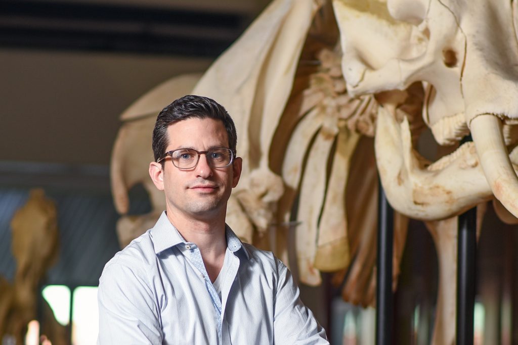
Alon Greenbaum
· Assistant Professor of Biomedical Engineering Affiliate Graduate Faculty (Bioinformatics)VerifiedNorth Carolina State University · Statistics
Active 2008–2025
Research signals
Five dimensions sourced from public faculty / publication signals. Sign in to compare against your own profile and see your match score.
Research topics
- Computer Science
- Biology
- Cell biology
- Biochemistry
- Chemistry
- Computational biology
- Genetics
Selected publications
UNC Libraries · 2025-05-22
erratumOpen accessSenior author[This corrects the article on p. 5214 in vol. 12, PMID: 34513252.].
Novel Porcine Model Reveals Two Distinct LGR5 Cell Types During Lung Development and Homeostasis.
UNC Libraries · 2025-11-06
articleOpen accessCells expressing LGR5 play a pivotal role in homeostasis, repair, and regeneration in multiple organs including skin and gastrointestinal tract, yet little is known about their role in the lung. Findings from mice, a widely used animal model, suggest that lung LGR5 expression differs from that of humans. In this work, using a new transgenic pig model, we identify two main populations of LGR5<sup>+</sup> cells in the lung that are conserved in human, but not mouse lungs. Using RNA sequencing, 3D imaging and organoid models, we determine that in the fetal lung, epithelial LGR5 expression is transient in a subpopulation of SOX9<sup>+</sup>/ETV<sup>+</sup>/SFTPC<sup>+</sup> progenitor lung tip cells. In contrast, epithelial LGR5 expression is absent from postnatal lung, but is reactivated in bronchioalveolar organoids derived from basal airway cells. We also describe a separate population of mesenchymal LGR5<sup>+</sup> cells that surrounds developing and mature airways, lies adjacent to airway basal cells, and is closely associated with nerve fibers. Transcriptionally, mesenchymal LGR5<sup>+</sup> cells include a subset of peribronchial fibroblasts (PBF) that express unique patterns of <em>SHH, FGF, WNT</em> and <em>TGF-β</em> signaling pathway genes. These results support distinct roles for LGR5<sup>+</sup> cells in the lung and describe a physiologically relevant animal model for further studies on the function of these cells in repair and regeneration.
Deep learning-based adaptive optics for light sheet fluorescence microscopy.
UNC Libraries · 2025-03-19
articleOpen accessSenior authorLight sheet fluorescence microscopy (LSFM) is a high-speed imaging technique that is often used to image intact tissue-cleared specimens with cellular or subcellular resolution. Like other optical imaging systems, LSFM suffers from sample-induced optical aberrations that decrement imaging quality. Optical aberrations become more severe when imaging a few millimeters deep into tissue-cleared specimens, complicating subsequent analyses. Adaptive optics are commonly used to correct sample-induced aberrations using a deformable mirror. However, routinely used sensorless adaptive optics techniques are slow, as they require multiple images of the same region of interest to iteratively estimate the aberrations. In addition to the fading of fluorescent signal, this is a major limitation as thousands of images are required to image a single intact organ even without adaptive optics. Thus, a fast and accurate aberration estimation method is needed. Here, we used deep-learning techniques to estimate sample-induced aberrations from only two images of the same region of interest in cleared tissues. We show that the application of correction using a deformable mirror greatly improves image quality. We also introduce a sampling technique that requires a minimum number of images to train the network. Two conceptually different network architectures are compared; one that shares convolutional features and another that estimates each aberration independently. Overall, we have presented an efficient way to correct aberrations in LSFM and to improve image quality.
UNC Libraries · 2025-04-26
articleOpen access1st authorCorrespondingLight-sheet fluorescence microscopy (LSFM) is a high-speed imaging technique that provides optical sectioning with reduced photodamage. LSFM is routinely used in life sciences for live cell imaging and for capturing large volumes of cleared tissues. LSFM has a unique configuration, in which the illumination and detection paths are separated and perpendicular to each other. As such, the image quality, especially at high resolution, largely depends on the degree of overlap between the detection focal plane and the illuminating beam. However, spatial heterogeneity within the sample, curved specimen boundaries, and mismatch of refractive index between tissues and immersion media can refract the well-aligned illumination beam. This refraction can cause extensive blur and non-uniform image quality over the imaged field-of-view. To address these issues, we tested a deep learning-based approach to estimate the angular error of the illumination beam relative to the detection focal plane. The illumination beam was then corrected using a pair of galvo scanners, and the correction significantly improved the image quality across the entire field-of-view. The angular estimation was based on calculating the defocus level on a pixel level within the image using two defocused images. Overall, our study provides a framework that can correct the angle of the light-sheet and improve the overall image quality in high-resolution LSFM 3D image acquisition.
Spiner, deep learning-based automated detection of spiral ganglion neurons in intact cochleae
iScience · 2025-06-18 · 1 citations
articleOpen accessSenior authorTissue clearing and light-sheet fluorescence microscopy were applied for 3D profiling of intact cochleae. However, the spiral ganglion neurons (SGNs) remain relatively understudied compared to hair cells and supporting cells, especially in large animal models. Here, we: (1) introduced collagenase treatment to the current protocol of tissue clearing to enhance uniform antibody staining of SGNs within the pig cochlea and (2) adopted a deep learning object detection model to locate and count SGNs in large 3D datasets via Spiner (Spiral ganglion neuron profiler). Using this approach, Type I SGNs in intact gerbil and pig cochleae were detected and counted in 3D, and Spiner counts were consistent with manual counts. We believe broad adaptation of the method will improve understanding of the SGN population and their role in hearing loss. Codes for a user-friendly web interface were provided for model running and fine-tuning, making it accessible to those without programming experience.
UNC Libraries · 2025-01-23
articleOpen accessNovel Porcine Model Reveals Two Distinct LGR5 Cell Types during Lung Development and Homeostasis
American Journal of Respiratory Cell and Molecular Biology · 2024-11-05 · 1 citations
articleOpen accessAbstract Cells expressing leucine-rich repeat-containing G-protein-coupled receptor 5 (LGR5) play a pivotal role in homeostasis, repair, and regeneration in multiple organs, including skin and gastrointestinal tract, but little is known about their role in the lung. Findings from mice, a widely used animal model, suggest that lung LGR5 expression differs from that of humans. In this work, using a new transgenic pig model, we identify two main populations of LGR5+ cells in the lung that are conserved in human but not mouse lungs. Using RNA sequencing, three-dimensional imaging, and organoid models, we determine that in the fetal lung, epithelial LGR5 expression is transient in a subpopulation of SOX9+/ETV5+/SFTPC+ progenitor lung tip cells. In contrast, epithelial LGR5 expression is absent from postnatal lung but is reactivated in bronchioalveolar organoids derived from basal airway cells. We also describe a separate population of mesenchymal LGR5+ cells that surrounds developing and mature airways, lies adjacent to airway basal cells, and is closely associated with nerve fibers. Transcriptionally, mesenchymal LGR5+ cells include a subset of peribronchial fibroblasts that express unique patterns of SHH, FGF, WNT, and TGF-β signaling pathway genes. These results support distinct roles for LGR5+ cells in the lung and describe a physiologically relevant animal model for further studies on the function of these cells in repair and regeneration.
Spiner, Deep Learning-Based Automated Detection of Spiral Ganglion Neurons in Intact Cochleae
SSRN Electronic Journal · 2024-01-01
preprintOpen accessSenior authorRoadmap on computational methods in optical imaging and holography [invited]
Applied Physics B · 2024-08-29 · 57 citations
reviewOpen accessComputational methods have been established as cornerstones in optical imaging and holography in recent years. Every year, the dependence of optical imaging and holography on computational methods is increasing significantly to the extent that optical methods and components are being completely and efficiently replaced with computational methods at low cost. This roadmap reviews the current scenario in four major areas namely incoherent digital holography, quantitative phase imaging, imaging through scattering layers, and super-resolution imaging. In addition to registering the perspectives of the modern-day architects of the above research areas, the roadmap also reports some of the latest studies on the topic. Computational codes and pseudocodes are presented for computational methods in a plug-and-play fashion for readers to not only read and understand but also practice the latest algorithms with their data. We believe that this roadmap will be a valuable tool for analyzing the current trends in computational methods to predict and prepare the future of computational methods in optical imaging and holography. Supplementary Information: The online version contains supplementary material available at 10.1007/s00340-024-08280-3.
Intelligent Beam Optimization for Light-Sheet Fluorescence Microscopy through Deep Learning
UNC Libraries · 2024-10-08
articleOpen accessLight-sheet fluorescence microscopy (LSFM) provides the benefit of optical sectioning coupled with rapid acquisition times, enabling high-resolution 3-dimensional imaging of large tissue-cleared samples. Inherent to LSFM, the quality of the imaging heavily relies on the characteristics of the illumination beam, which only illuminates a thin section of the sample. Therefore, substantial efforts are dedicated to identifying slender, nondiffracting beam profiles that yield uniform and high-contrast images. An ongoing debate concerns the identification of optimal illumination beams for different samples: Gaussian, Bessel, Airy patterns, and/or others. However, comparisons among different beam profiles are challenging as their optimization objectives are often different. Given that our large imaging datasets (approximately 0.5 TB of images per sample) are already analyzed using deep learning models, we envisioned a different approach to the problem by designing an illumination beam tailored to boost the performance of the deep learning model. We hypothesized that integrating the physical LSFM illumination model (after passing it through a variable phase mask) into the training of a cell detection network would achieve this goal. Here, we report that joint optimization continuously updates the phase mask and results in improved image quality for better cell detection. The efficacy of our method is demonstrated through both simulations and experiments that reveal substantial enhancements in imaging quality compared to the traditional Gaussian light sheet. We discuss how designing microscopy systems through a computational approach provides novel insights for advancing optical design that relies on deep learning models for the analysis of imaging datasets.
Recent grants
Frequent coauthors
- 83 shared
Adele Moatti
UNC/NCSU Joint Department of Biomedical Engineering
- 82 shared
Yuheng Cai
- 80 shared
Aydogan Özcan
University of California, Los Angeles
- 38 shared
Wei Luo
University of California, Los Angeles
- 38 shared
Frances S. Ligler
Texas A&M University
- 32 shared
Mani Ratnam
- 29 shared
Dylan Silkstone
North Carolina State University
- 25 shared
Chen Li
Weatherford College
- Resume-aware match score
- Save to shortlist
- AI-drafted outreach
See your match with Alon Greenbaum
PhdFit ranks faculty by your research interests, methods, and publications — grounded in their actual work, not templates.
- Free to start
- No credit card
- 30-second signup