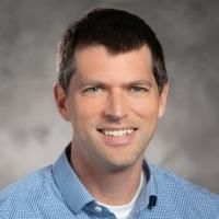
Donald T Fox
· Professor of Pharmacology and Cancer BiologyVerifiedDuke University · Genetics and Genomics
Active 2001–2025
About
Donald T Fox is a Professor of Pharmacology and Cancer Biology and a Professor of Cell Biology at Duke University School of Medicine. He is a member of the Duke Cancer Institute and an associate of the Duke Initiative for Science & Society. Additionally, he is affiliated with the Duke Regeneration Center. His academic roles are supported by his involvement in various programs including the Program in Cell and Molecular Biology, the Developmental & Stem Cell Biology Program, and the Third Year University Program in Genetics and Genomics. His work is situated within Duke's broader research and educational efforts, contributing to the fields of pharmacology, cancer biology, cell biology, and regenerative sciences.
Research topics
- Biology
- Cell biology
- Genetics
- Evolutionary biology
- Computational biology
- Ecology
- Neuroscience
- Anatomy
- Endocrinology
- Physiology
Selected publications
Spatial ploidy inference using quantitative imaging
bioRxiv (Cold Spring Harbor Laboratory) · 2025-03-13 · 1 citations
preprintOpen accessPolyploidy (whole-genome multiplication) is a common yet under-surveyed property of tissues across multicellular organisms. Polyploidy plays a critical role during tissue development, following acute stress, and during disease progression. Common methods to reveal polyploidy involve either destroying tissue architecture by cell isolation or by tedious identification of individual nuclei in intact tissue. Therefore, there is a critical need for rapid and high-throughput ploidy quantification using images of nuclei in intact tissues. Here, we present iSPy (Inferring Spatial Ploidy), a new unsupervised learning pipeline that is designed to create a spatial map of nuclear ploidy across a tissue of interest. We demonstrate the use of iSPy in Arabidopsis, Drosophila, and human tissue. iSPy can be adapted for a variety of tissue preparations, including whole mount and sectioned. This high-throughput pipeline will facilitate rapid and sensitive identification of nuclear ploidy in diverse biological contexts and organisms.
Spatial ploidy inference using quantitative imaging
Cell Reports Methods · 2025-12-01
articleOpen accessPolyploidy (whole-genome duplication) is a common yet under-surveyed property of tissues across multicellular organisms. Polyploidy plays a critical role during tissue development, following acute stress, and during disease progression. Common methods to reveal polyploidy involve either destroying tissue architecture by cell isolation or tedious identification of individual nuclei in intact tissue. Therefore, there is a critical need for rapid and high-throughput ploidy quantification using images of nuclei in intact tissues. Here, we present iSPy (inferring Spatial Ploidy), an unsupervised learning pipeline that is designed to create a spatial map of nuclear ploidy across a tissue of interest. We demonstrate the use of iSPy in Arabidopsis, Drosophila, and human tissue. iSPy can be adapted for a variety of tissue preparations, including whole mount and sectioned. This high-throughput pipeline will facilitate rapid and sensitive identification of nuclear ploidy in diverse biological contexts and organisms.
Synaptic vesicle glycoprotein 2 enables viable aneuploidy following centrosome amplification
bioRxiv (Cold Spring Harbor Laboratory) · 2025-02-20
preprintOpen accessSenior authorCorrespondingAbstract Amplified centrosome number causes genomic instability, most severely through division into more than two aneuploid daughter cells (multipolar mitosis). Several mechanisms that suppress multipolar division have been uncovered, yet mechanisms that favor viable multipolar division are poorly understood. To uncover factors that promote viability in cells with frequent centrosome amplification and multipolar division, we conducted an unbiased Drosophila genetic screen. In 642 mutagenized lines, we exploited the ability of intestinal papillar cells to form and function despite multipolar divisions. Our top hit is an unnamed gene, CG3168 . We name this gene synaptic vesicle glycoprotein 2 , reflecting homology to human Synaptic Vesicle Glycoprotein 2 (SV2) proteins. GFP-tagged SV2 localizes to the plasma membrane. In cells with amplified centrosomes, SV2 positions membrane-adjacent centrosomes, which prevents severe errors in chromosome alignment and segregation. Our results uncover membrane-based multipolar division regulation and reveal a novel vulnerability in cells with common cancer properties.
High-speed 4D fluorescence light field tomography of whole freely moving organisms
Optica · 2025-04-04 · 6 citations
articleOpen accessVolumetric fluorescence imaging techniques, such as confocal, multiphoton, light sheet, and light field microscopy, have become indispensable tools across a wide range of cellular, developmental, and neurobiological applications. However, it is difficult to scale such techniques to the large 3D fields of view (FOV), volume rates, and synchronicity requirements for high-resolution 4D imaging of freely behaving organisms. Here, we present reflective Fourier light field computed tomography (ReFLeCT), a high-speed volumetric fluorescence computational imaging technique. ReFLeCT synchronously captures entire tomograms of multiple unrestrained, unanesthetized model organisms across multi-millimeter 3D FOVs at 120 volumes per second. In particular, we applied ReFLeCT to reconstruct 4D videos of fluorescently labeled zebrafish and Drosophila larvae, enabling us to study their heartbeat, fin and tail motion, gaze, jaw motion, and muscle contractions with nearly isotropic 3D resolution while they are freely moving. To our knowledge, as a novel approach for snapshot tomographic capture, ReFLeCT is a major advance toward bridging the gap between current volumetric fluorescence microscopy techniques and macroscopic behavioral imaging.
bioRxiv (Cold Spring Harbor Laboratory) · 2025-04-05
preprintOpen accessSenior authorCorrespondingAbstract Tissue maintenance in the presence of cell death-promoting insults requires a host of molecular mechanisms. Many studies focus on cell renewal through regeneration, while fewer studies explore mechanisms that promote cell longevity despite cell death stimuli. Here, we reveal that the adult Drosophila hindgut ileum is an excellent model to study tissue maintenance by long-lived cells. Hindgut ileal enterocytes resist the damaging detergent SDS and upstream caspase signaling by head-involution-defective (hid). This hid- induced death insensitivity arises early in adulthood and associates with numerous transcriptional changes. We interrogated 82 of these transcriptional changes in a candidate screen for enhancers of hid- induced death in the ileum. Top among our screen hits is an immunoglobulin family cell adhesion gene, CG15312. CG15312 maintains the adhesion protein FasIII on cell membranes. In hid- expressing ileal cells, CG15312 loss causes cell death and pyknotic nuclear clustering. We name this conserved gene lo w o n-membrane f a s and enhancer of h id ( loofah ). Our findings reveal a new mechanism linking cell adhesion and cell death resistance in a long-lived cell type. Our work establishes a new model to study tissue preservation.
Organ-specific rewiring of mitochondrial integrity through COX7A dictates cellular ploidy control
bioRxiv (Cold Spring Harbor Laboratory) · 2025-09-30 · 1 citations
preprintOpen accessSenior authorCorrespondingTo achieve proper cell and tissue size, cytoplasmic and nuclear growth must be coordinated. Disrupting this coordination causes birth defects and disease. In nature's largest cells, nuclear growth occurs through polyploidization (whole-genome-duplication). How the massive nuclear growth of polyploid cells is coordinated with cytoplasmic growth processes such as mitochondrial biogenesis is relatively unclear. Here, focusing on one of nature's most commonly polyploid organs-the heart-we uncover cross-talk between cytoplasmic mitochondrial biogenesis/integrity and nuclear growth/polyploidy. From a human-to-fly screen, we uncover novel regulators of cardiomyocyte ploidy, including mitochondrial integrity regulators. In comparing these cardiac hits with a parallel screen in another polyploid tissue, the salivary gland, we discovered two opposing roles for Cytochrome-c-oxidase-subunit-7A (COX7A). While salivary gland COX7A preserves mitochondrial integrity to promote polyploidy and optimal organ growth, cardiac COX7A instead suppresses mitochondrial biogenesis to repress polyploidy and prevent hypertrophic organ growth. Among all electron transport chain genes, only COX7A functions as a cardiac growth repressor. Fly hearts with compromised COX7A show abnormally high cardiac output. Human COX7A1, a mitochondrial-localized protein, similarly represses polyploidy of human iPSC-derived cardiomyocytes. In summary, our human-fly-human approach reveals conserved rewiring of mitochondrial integrity in heart tissue that switches COX7A's role from ploidy promotion to repression. Our findings reveal fundamental cross-talk between mitochondrial biogenesis and genome duplication that are critical in growing metazoan tissues.
Synaptic vesicle glycoprotein 2 enables viable aneuploidy following centrosome amplification
Genetics · 2025-07-31
articleOpen accessSenior authorAmplified centrosome number causes genomic instability, most severely through division into >2 aneuploid daughter cells (multipolar mitosis). Several mechanisms that suppress multipolar division have been uncovered, yet mechanisms that favor viable multipolar division are poorly understood. To uncover factors that promote viability in cells with frequent centrosome amplification and multipolar division, we conducted an unbiased Drosophila genetic screen. In 642 mutagenized lines, we exploited the ability of intestinal papillar cells to form and function despite multipolar divisions. Our top hit is an unnamed gene, CG3168. We name this gene synaptic vesicle glycoprotein 2, reflecting homology to human Synaptic Vesicle Glycoprotein 2 (SV2) proteins. GFP-tagged SV2 localizes to the plasma membrane. In cells with amplified centrosomes, SV2 positions membrane-adjacent centrosomes, which prevents severe errors in chromosome alignment and segregation. Our results uncover membrane-based multipolar division regulation and reveal a novel vulnerability in cells with common cancer properties.
Loofah suppresses cell death in long-lived <i>Drosophila</i> hindgut enterocytes
Development · 2025-12-15
articleOpen accessSenior authorTissue maintenance in the presence of cell death-promoting insults requires a host of molecular mechanisms. Many studies focus on cell renewal through regeneration, while fewer studies explore mechanisms that promote cell longevity despite cell death stimuli. Here, we reveal that the adult Drosophila hindgut ileum is an excellent model for studying tissue maintenance by long-lived cells. Hindgut ileal enterocytes resist the damaging detergent SDS and upstream caspase signaling by head-involution-defective (hid). This hid-induced death insensitivity arises early in adulthood and is associated with numerous transcriptional changes. We interrogated 82 of these transcriptional changes in a candidate screen for enhancers of hid-induced death in the ileum. Top among our screen hits is an immunoglobulin family cell adhesion gene, CG15312, that maintains the adhesion protein FasIII on cell membranes. In hid-expressing ileal cells, CG15312 loss causes cell death and pyknotic nuclear clustering. We name this conserved gene low on-membrane fas and enhancer of hid (loofah). Our findings reveal a new mechanism linking cell adhesion and cell death resistance in a long-lived cell type. Our work establishes a new model for studying tissue preservation.
2024-03-12
articleIt is challenging to study behavior of and track freely-moving model organisms using conventional 3D microscopy techniques. To overcome motion artifacts and prevent the organism from leaving the field of view (FOV), existing techniques require paralyzing or otherwise immobilizing the organism. Here, we demonstrate hemispheric Fourier light field tomography, featuring a parabolic objective that enables synchronized multi-view fluorescence imaging over ~2pi steradians at up to 120 fps and across multi-millimeter 3D FOVs. Our method is not only able to track the 6D pose of freely-moving zebrafish and fruit fly larvae, but also other properties such as heartbeat, fin motion, jaw motion, and muscle contractions. We also demonstrate simultaneous multi-organism imaging.
Orb2 enables rare-codon-enriched mRNA expression during Drosophila neuron differentiation
Nature Communications · 2024-06-20 · 9 citations
articleOpen accessSenior authorRegulation of codon optimality is an increasingly appreciated layer of cell- and tissue-specific protein expression control. Here, we use codon-modified reporters to show that differentiation of Drosophila neural stem cells into neurons enables protein expression from rare-codon-enriched genes. From a candidate screen, we identify the cytoplasmic polyadenylation element binding (CPEB) protein Orb2 as a positive regulator of rare-codon-dependent mRNA stability in neurons. Using RNA sequencing, we reveal that Orb2-upregulated mRNAs in the brain with abundant Orb2 binding sites have a rare-codon bias. From these Orb2-regulated mRNAs, we demonstrate that rare-codon enrichment is important for mRNA stability and social behavior function of the metabotropic glutamate receptor (mGluR). Our findings reveal a molecular mechanism by which neural stem cell differentiation shifts genetic code regulation to enable critical mRNA stability and protein expression.
Recent grants
Polyploidy after tissue injury: a Drosophila model
NIH · $3.1M · 2016–2026
Frequent coauthors
- 42 shared
Benjamin M. Stormo
Duke University
- 30 shared
Erez Cohen
University of Michigan–Ann Arbor
- 25 shared
Nora G. Peterson
Duke University
- 24 shared
Jessica K. Sawyer
- 22 shared
Allan C. Spradling
Carnegie Institution for Science
- 16 shared
Pamela S. Soltis
Florida Museum of Natural History
- 14 shared
Juliet S. King
Duke University
- 14 shared
Scott R. Allen
University of North Carolina at Chapel Hill
Education
- 2006
PhD, Biology
University of North Carolina
- Resume-aware match score
- Save to shortlist
- AI-drafted outreach
See your match with Donald T Fox
PhdFit ranks faculty by your research interests, methods, and publications — grounded in their actual work, not templates.
- Free to start
- No credit card
- 30-second signup